

Former Fellow on Team That Surmounted X-ray Imaging Obstacle
A Department of Energy Computational Science Graduate Fellowship (DOE CSGF) alumnus is part of an international research team that developed algorithms cracking a longstanding barrier to capturing the three-dimensional structures of tiny biological objects.
Lawrence Berkeley National Laboratory researchers, including former fellow Jeffrey Donatelli, devised a mathematical scheme that analyzes data from powerful X-ray imaging tools. Their paper, published Oct. 12 in the journal Physical Review Letters, tells how the technique revealed the structural features, measured in billionths of a meter, of viruses. The information could reveal valuable insights into disease and other biological processes.
Donatelli and colleagues Peter Zwart and Kanupriya Pande are part of Berkeley Lab’s Center for Advanced Mathematics for Energy Research Applications (CAMERA). They focused on single-particle X-ray diffraction experiments conducted at DOE’s Linac Coherent Light Source (LCLS) at SLAC National Accelerator Laboratory.
The LCLS X-ray free electron laser (XFEL) generates bright, short X-ray pulses that diffract off of single particles. The diffraction patterns, captured by sensitive detectors, provide clues to the objects’ structures.
The problem: It’s unclear from the diffraction data how the single particles are oriented – which direction they’re turned in relation to the laser. “You don’t have control over rotation of the particles as they are hit by the X-ray beam, so each snapshot from a successful hit will contain information about the sample from a different orientation,” Donatelli said in a Berkeley Lab release. Scientists try to determine orientation from the images themselves, but noisy data makes that difficult.
Rather than directly determine the orientation, the team’s technique relies on analyzing the angular correlations of bright, ultrashort XFEL beams as they scatter off the particle. The correlation data “contains a comprehensive fingerprint of the 3-D structure of an object that extends traditional solution scattering approaches,” Zwart said.
CAMERA devised a multi-tiered iterative phasing (M-TIP) algorithm that solves the structure “directly from the correlation data without having to rely on any symmetry constraints, and, more importantly, without the need to solve the orientation determination problem,” Donatelli said.
Donatelli was in the DOE CSGF from 2009 to 2013, when he earned a doctorate in applied mathematics from the University of California, Berkeley.
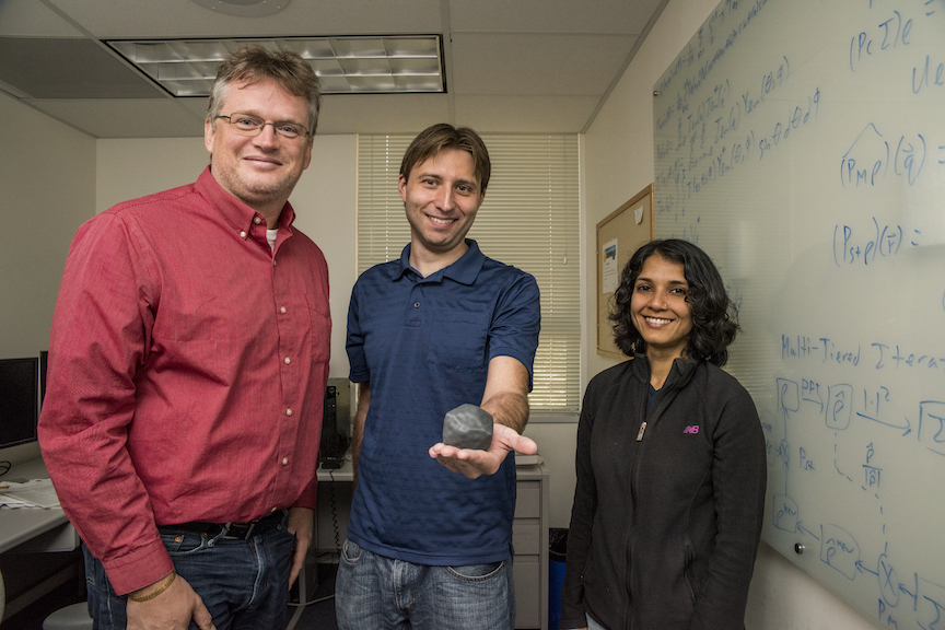
Image caption: CAMERA members (from left) Peter Zwart, Jeffrey Donatelli and Kanupriya Pande, co-authors of a paper describing how the group’s M-TIP algorithm determined 3-D virus structures from single-particle diffraction data. Donatelli holds a 3-D-printed model of one of the viruses M-TIP reconstructed. Credit: Marilyn Chung, Berkeley Lab.